
ملف:Human brain anterior-inferior view description.JPG
Human_brain_anterior-inferior_view_description.JPG (330 × 475 بكسل حجم الملف: 31 كيلوبايت، نوع MIME: image/jpeg)
| هذا ملف من ويكيميديا كومنز. معلومات من صفحة وصفه مبينة في الأسفل. كومنز مستودع ملفات ميديا ذو رخصة حرة. |
ملخص
| الوصف |
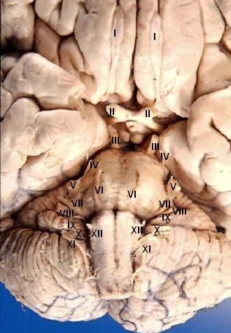
Human brain - anterior-inferior view - cerebral nerves I. N. olfactorius - The fila olfactoria (approximately 20 nerve rootlets on each side) comprise the Olfactory nerves (I) which run from the olfactory receptors in the nasal cavity to the olfactory bulb. II. N. opticus - The Optic nerves (II) are large & join at the midline to form the optic chiasm, then continue laterally as the optic tracts. The optic nerve is not a true nerve but rather a CNS tract. III. N. oculomotorius - The Oculomotor nerves (III) emerge from a depression in the midbrain, the interpeduncular fossa, just caudal to the optic chiasm. IV. N. trochlearis - The Trochlear nerves (IV) are small & are seen on the lateral surface of the midbrain. They are the only nerves which exit from the posterior side of the brain. V. N. trigeminus - The Trigeminal nerves (V) are large and emanate from the lateral surface of the pons. VI. N. abducens - The Abducens nerves (VI) exit near the midline from the inferior pontine sulcus which separates the pons from the medulla. VII. N. facialis - Moving laterally in the inferior pontine sulcus, the Facial nerves (VII) can be seen. VIII. N. vestibulocochlearis - Slightly lateral to the facial nerve is the Vestibulocochlear nerve (VIII). IX. N. glossopharyngeus - The postolivary sulcus is a groove running rostrocaudally on the lateral surface of the medulla. From this sulcus pass the small Glossopharyngeal nerves (IX) rostrally and... X. N. vagus - ...the much larger Vagus nerves (X) caudally. XI. N. accessorius - The Spinal Accessory nerves (XI) exit the cervical cord then pass rostrally through the foramen magnum to exit the cranial vault with the lossopharyngeal and vagus nerves. XII. N. hypoglossus - The Hypoglossal nerves (XII) exit the medulla via the preolivary sulcus.
|
| التاريخ | |
| المصدر | http://www.healcentral.org/healapp/showMetadata?metadataId=40566 (Internet Archive of file description page) |
| المؤلف |
John A Beal, PhD Dep't. of Cellular Biology & Anatomy, Louisiana State University Health Sciences Center Shreveport |
| الترخيص (إعادة استخدام هذا الملف) |
CC-BY |
| إصدارات أخرى |
 |
ترخيص
- يحقُّ لك:
- مشاركة العمل – نسخ العمل وتوزيعه وبثُّه
- إعادة إنتاج العمل – تعديل العمل
- حسب الشروط التالية:
- نسب العمل إلى مُؤَلِّفه – يلزم نسب العمل إلى مُؤَلِّفه بشكل مناسب وتوفير رابط للرخصة وتحديد ما إذا أجريت تغييرات. بالإمكان القيام بذلك بأية طريقة معقولة، ولكن ليس بأية طريقة تشير إلى أن المرخِّص يوافقك على الاستعمال.
رُوجِع هذا الملفُّ الذي نشر أوَّلاً في الموقع http://www.healcentral.org/healapp/showMetadata?metadataId=40566، بتاريخ 25 September 2013 من قبل المُراجِع Eleassar الذي أكَّد
أنَّه كان مُتوافِراً في ذلك المَوقع في ذلك التاريخ تحت الرُّخصة المُشار إِليها.
|
تاريخ الملف
اضغط على زمن/تاريخ لرؤية الملف كما بدا في هذا الزمن.
| زمن/تاريخ | صورة مصغرة | الأبعاد | مستخدم | تعليق | |
|---|---|---|---|---|---|
| حالي | 18:22، 24 يونيو 2006 | | 330 × 475 (31 كيلوبايت) | Patho | {{Information| |Description='''Human brain - anterior-inferior view - cerebral nerves''' I. N. olfactorius - The fila olfactoria (approximately 20 nerve rootlets on each side) comprise the Olfactory nerves (I) which run from the olfactory receptors in |
استخدام الملف
الصفحة التالية تستخدم هذا الملف:
الاستخدام العالمي للملف
الويكيات الأخرى التالية تستخدم هذا الملف:
- الاستخدام في azb.wikipedia.org
- الاستخدام في az.wikipedia.org
- الاستخدام في bs.wikipedia.org
- الاستخدام في de.wikipedia.org
- الاستخدام في de.wikibooks.org
- الاستخدام في en.wikipedia.org
- Cranial nerves
- User talk:Hovea
- User talk:Wouterstomp
- User talk:Nephron
- User talk:NCurse
- User talk:Robotsintrouble
- User talk:Was a bee
- User talk:Bloomingdedalus
- User talk:Read-write-services
- User talk:Attys
- User talk:Bakerstmd
- User talk:Manfi
- User talk:Mikepascoe
- User talk:Vokesk
- User talk:Neuraxıs
- User talk:Cmungall
- User talk:Qxukhgiels
- User talk:DocElisa
- User talk:JakobSteenberg/Archives/1
- User talk:Meteor sandwich yum
- User talk:Slashedone
- User talk:Anindya07
- User talk:Mattimussi
- User talk:Jelly Bean MD
- User talk:Benrusholme
- User talk:Brad.w.english
- User talk:Krsna ss
- User talk:Tom (LT)/Archive 4
- User talk:Dregon131
- User talk:Sapientia42
- User talk:Tilifa Ocaufa
- User talk:Snow Rise/Archive 6
- User talk:Arcadian
- User talk:Outofbattery
- User talk:Anatomyczar
- Wikipedia:WikiProject Anatomy/Newsletter/4
- User talk:Supravibhatsupravi
- User talk:Athikhun.suw
- User talk:CFCF/Archive 6
- User talk:Lewisskinner/ArchiveJul 2015
- User talk:Tyrol5/Archive 6
- User talk:CFCF/Archive 7
- User talk:DiverDave/Archive 5
- User talk:Caledones/archive/archive4
اعرض المزيد من الاستخدام العام لهذا الملف.