
ملف:6nb6 prefusion 6m3w postfusion spike.png
الملف الأصلي (768 × 1٬536 بكسل حجم الملف: 382 كيلوبايت، نوع MIME: image/png)
| هذا ملف من ويكيميديا كومنز. معلومات من صفحة وصفه مبينة في الأسفل. كومنز مستودع ملفات ميديا ذو رخصة حرة. |
ملخص
| الوصف |
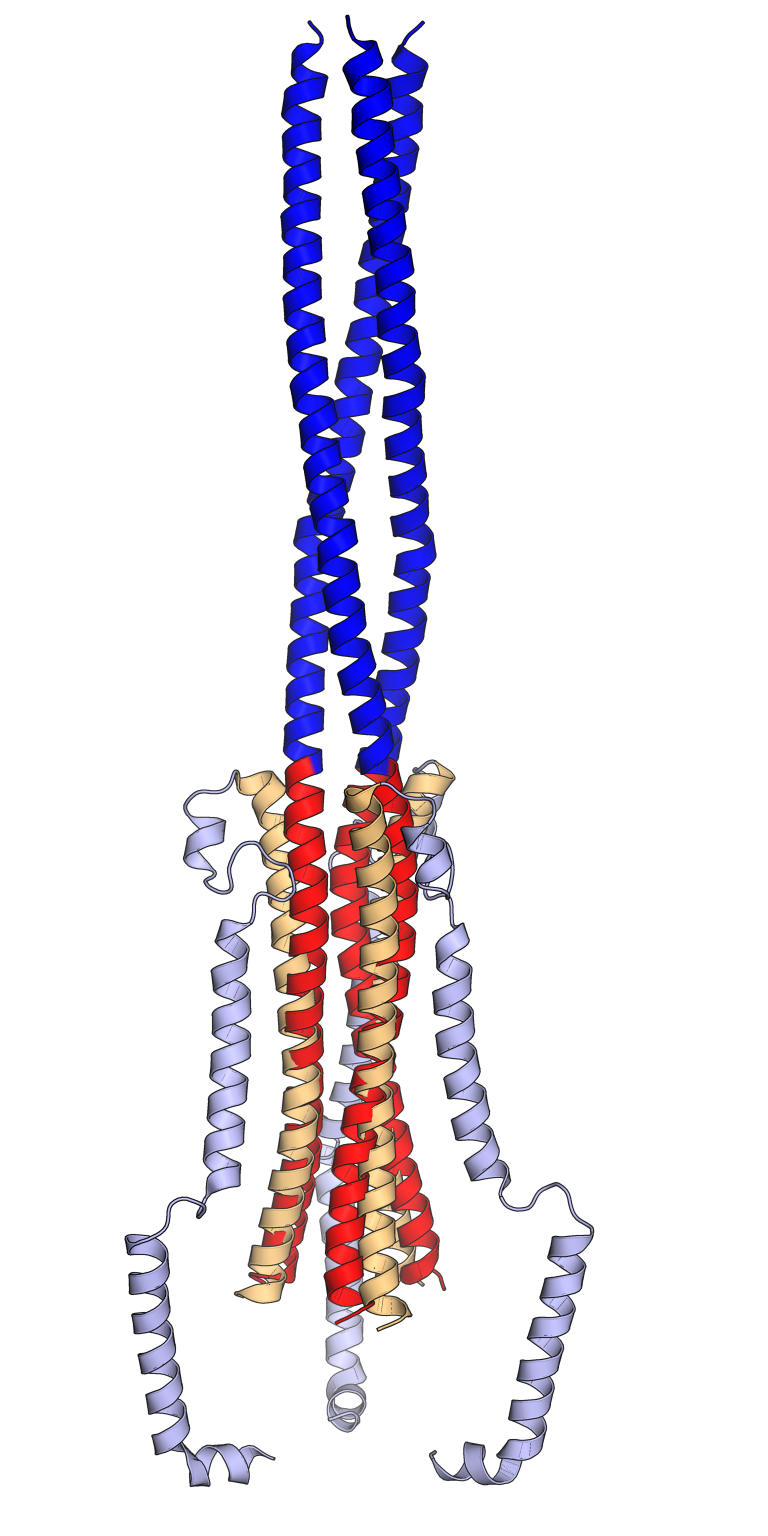
Comparison of the pre-fusion (orange, light blue) and post-fusion (red, dark blue) conformations of the SARS-CoV spike protein trimer. In the pre-fusion conformation, the central helix (orange) and heptad repeat 1 (HR1, light blue) are folded back on each other in an antiparallel orientation. In the post-fusion conformation, the central helix (red) and the HR1 sequence (dark blue) reorganize to form an extended trimeric coiled coil. The viral membrane is at the bottom and the host cell membrane at the top. Only key portions of the S2 subunit are shown. Rendered using PyMol from cryo-electron microscopy structures PDB: 6NB6 (pre-fusion) and PDB: 6M3W (post-fusion) superposed using the central helix sequences, inspired by Figs 1 and 2 from Fan 2020. 6NB6: Unexpected Receptor Functional Mimicry Elucidates Activation of Coronavirus Fusion. Walls, A.C., Xiong, X., Park, Y.J., Tortorici, M.A., Snijder, J., Quispe, J., Cameroni, E., Gopal, R., Dai, M., Lanzavecchia, A., Zambon, M., Rey, F.A., Corti, D., Veesler, D. (2019) Cell 176: 1026-1039.e15 PubMed: 30712865 DOI: 10.1016/j.cell.2018.12.028 6M3W: Cryo-EM analysis of the post-fusion structure of the SARS-CoV spike glycoprotein. Fan, X., Cao, D., Kong, L., Zhang, X. (2020) Nat Commun 11: 3618-3618 PubMed: 32681106 DOI: 10.1038/s41467-020-17371-6 |
| التاريخ | |
| المصدر | عمل شخصي |
| المؤلف | Opabinia regalis |
ترخيص
- يحقُّ لك:
- مشاركة العمل – نسخ العمل وتوزيعه وبثُّه
- إعادة إنتاج العمل – تعديل العمل
- حسب الشروط التالية:
- نسب العمل إلى مُؤَلِّفه – يلزم نسب العمل إلى مُؤَلِّفه بشكل مناسب وتوفير رابط للرخصة وتحديد ما إذا أجريت تغييرات. بالإمكان القيام بذلك بأية طريقة معقولة، ولكن ليس بأية طريقة تشير إلى أن المرخِّص يوافقك على الاستعمال.
- الإلزام بترخيص المُشتقات بالمثل – إذا أعدت إنتاج المواد أو غيرت فيها، فيلزم أن تنشر مساهماتك المُشتقَّة عن الأصل تحت ترخيص الأصل نفسه أو تحت ترخيص مُتوافِقٍ معه.

|
يسمح نسخ وتوزيع و/أو تعديل هذه الوثيقة تحت شروط رخصة جنو للوثائق الحرة، الإصدار 1.2 أو أي إصدار لاحق تنشره مؤسسة البرمجيات الحرة؛ دون أقسام ثابتة ودون نصوص أغلفة أمامية ودون نصوص أغلفة خلفية. نسخة من الرخصة تم تضمينها في القسم المسمى GNU Free Documentation License. |
تاريخ الملف
اضغط على زمن/تاريخ لرؤية الملف كما بدا في هذا الزمن.
| زمن/تاريخ | صورة مصغرة | الأبعاد | مستخدم | تعليق | |
|---|---|---|---|---|---|
| حالي | 07:02، 13 سبتمبر 2021 | | 768 × 1٬536 (382 كيلوبايت) | Opabinia regalis | {{Information |Description=Comparison of the pre-fusion (orange, light blue) and post-fusion (red, dark blue) conformations of the SARS-CoV spike protein trimer. In the pre-fusion conformation, the central helix (orange) and heptad repeat 1 (HR1, light blue) are folded back on each other in an antiparallel orientation. In the post-fusion conformation, the central helix (red) and the HR1 sequence (dark blue) reorganize to form an extended trimeric coiled coil. The viral membrane is at the bott... |
استخدام الملف
الصفحة التالية تستخدم هذا الملف:
الاستخدام العالمي للملف
الويكيات الأخرى التالية تستخدم هذا الملف:
- الاستخدام في ca.wikipedia.org
- الاستخدام في de.wikipedia.org
- الاستخدام في en.wikipedia.org
- الاستخدام في es.wikipedia.org