
ملف:Bundleofhis.png
Bundleofhis.png (400 × 483 بكسل حجم الملف: 69 كيلوبايت، نوع MIME: image/png)
| هذا ملف من ويكيميديا كومنز. معلومات من صفحة وصفه مبينة في الأسفل. كومنز مستودع ملفات ميديا ذو رخصة حرة. |
ملخص
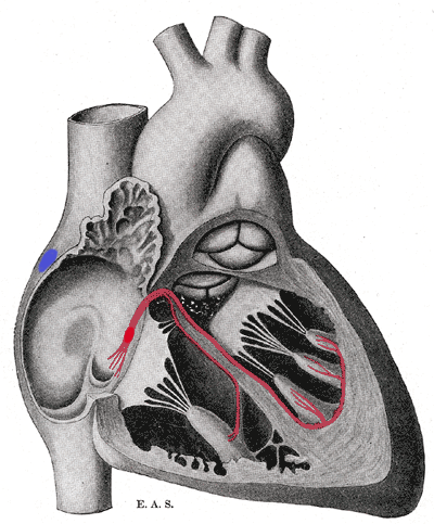
Schematic representation of the atrioventricular bundle of His. The bundle, represented in red, originates near the orifice of the coronary sinus, undergoes slight enlargement to form the AV node. The AV node tapers down into the bundle of HIS, which passes into the ventricular septum and divides into two bundle branches, the left and right bundles. Sometimes the 'left and right bundles of His' are called Purkyne or Purkinge fibres. The ultimate distribution cannot be completely shown in this diagram.
This image is misleading. Although it correctly places the SA and AV nodes in the right atrium, it appears as though there are only two papillary muscles in the right ventricle and three in the left ventricle. The opposite is actually true. The papillary muscles attach to chordae tendinae which then attach to the leaflets of the AV valves, preventing prolapse. The left AV valve is the mitral or bicuspid and only has two leaflets and therefore two papillary muscles. The right AV valve is the tricuspid and should have three papillary muscles corresponding to the three leaflets of the valve.
ترخيص
يقع ملفُ الوسائط هذا في النِّطاق العامّ داخل الولايات المُتحدة الأمريكيَّة. وهذا ينطبق على الأعمال أمريكيَّة الأصل التي نفدت مدة حقوق تأليفها ونشرها، وهذا في الغالب الأعم عائدٌ إلى نشرها للمرة الأولى مرة قبل 1 يناير، ١٩٢٩. راجع هذه الصفحة للمزيد من التوضيح.
|
 | |
 |
قد لا تكون هذه الصورة في النِّطاق العامّ من خارج الولايات المُتحدة، وهذا ينطبق بشكلٍ خاصٍّ في البلدان والمناطق التي لا يكون حكم الفترة الأقصر فيها نافذاً على الأعمال الأمريكية، مثل كندا وجمهورية الصين (ما خلا هونغ كونغ وماكاو) وألمانيا والمكسيك وسويسرا. إنَّ اسم المُؤَلِّف وسنة النشر هما من المعلومات الأساسيَّة، ولا بدَ من ذكرهما. انظر ويكيبيديا:الملكية العامة وويكيبيديا:حقوق التأليف والنشر للمزيد من التفاصيل.
|
تاريخ الملف
اضغط على زمن/تاريخ لرؤية الملف كما بدا في هذا الزمن.
| زمن/تاريخ | صورة مصغرة | الأبعاد | مستخدم | تعليق | |
|---|---|---|---|---|---|
| حالي | 17:40، 20 سبتمبر 2006 | | 400 × 483 (69 كيلوبايت) | Kauczuk | Bundle of His, from Gray's Anatomy 1918 |
استخدام الملف
الصفحة التالية تستخدم هذا الملف:
الاستخدام العالمي للملف
الويكيات الأخرى التالية تستخدم هذا الملف:
- الاستخدام في az.wikipedia.org
- الاستخدام في bn.wikibooks.org
- الاستخدام في bs.wikipedia.org
- الاستخدام في ca.wikipedia.org
- الاستخدام في cs.wikipedia.org
- الاستخدام في de.wikipedia.org
- الاستخدام في de.wikibooks.org
- الاستخدام في el.wikipedia.org
- الاستخدام في en.wikipedia.org
- الاستخدام في en.wikibooks.org
- الاستخدام في es.wikipedia.org
- الاستخدام في es.wikibooks.org
- الاستخدام في eu.wikipedia.org
- الاستخدام في fr.wikipedia.org
- الاستخدام في hi.wikipedia.org
- الاستخدام في it.wikipedia.org
- الاستخدام في ja.wikipedia.org
- الاستخدام في ja.wikibooks.org
- الاستخدام في ko.wikipedia.org
- الاستخدام في lv.wikipedia.org
- الاستخدام في nl.wikipedia.org
- الاستخدام في pl.wikipedia.org
- الاستخدام في pt.wikipedia.org
- الاستخدام في sr.wikipedia.org
- الاستخدام في www.wikidata.org