
ملف:HLA-A1.png
حجم هذه المعاينة: 539 × 599 بكسل. البعدان الآخران: 216 × 240 بكسل | 548 × 609 بكسل.
الملف الأصلي (548 × 609 بكسل حجم الملف: 145 كيلوبايت، نوع MIME: image/png)
| هذا ملف من ويكيميديا كومنز. معلومات من صفحة وصفه مبينة في الأسفل. كومنز مستودع ملفات ميديا ذو رخصة حرة. |
ملخص
| الوصف |
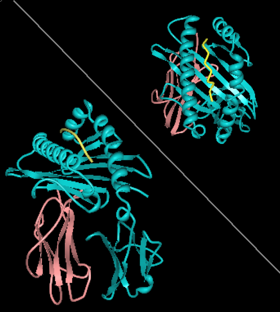
English: Rendering of HLA-A1 with MAGE-1 bound peptide. Two views, from the side showing B2 microglobulin (rose) and HLA-A1 (alpha chain, cyan). Top-right view is looking down through the binding site toward the plasma membrane. The image is derived from PDB:1W72 that was presented in the work:Hulsmeyer et al. (2005) A major histocompatibility complex-peptide-restricted antibody and t cell receptor molecules recognize their target by distinct binding modes: crystal structure of human leukocyte antigen (HLA)-A1-MAGE-A1 in complex with FAB-HYB3. J.Biol.Chem. 280: 2972-2980. Image rendered with PDB ProteinWorkshop 1.50. Several aspects of the image removed for clarity reasons. |
| المصدر | عمل شخصي |
| المؤلف | Pdeitiker |
ترخيص
| أنا، مالِك حقوق تأليف ونشر هذا العمل، أجعله في النِّطاق العامِّ، يسري هذا في أرجاء العالم كلِّه. في بعض البلدان، قد يكون هذا التَّرخيص غيرَ مُمكنٍ قانونيَّاً، في هذه الحالة: أمنح الجميع حق استخدام هذا العمل لأي غرض دون أي شرط ما لم يفرض القانون شروطًا إضافية. |
تاريخ الملف
اضغط على زمن/تاريخ لرؤية الملف كما بدا في هذا الزمن.
| زمن/تاريخ | صورة مصغرة | الأبعاد | مستخدم | تعليق | |
|---|---|---|---|---|---|
| حالي | 20:21، 21 أغسطس 2008 | | 548 × 609 (145 كيلوبايت) | Pdeitiker | {{Information |Description={{en|1=Rendering of HLA-A1 with MAGE-1 bound peptide. Two views, from the side showing B2 microglobulin (rose) and HLA-A1 (alpha chain, cyan). Top-right view is looking down through the binding site toward the plasma membrane. T |
استخدام الملف
الصفحة التالية تستخدم هذا الملف:
الاستخدام العالمي للملف
الويكيات الأخرى التالية تستخدم هذا الملف:
- الاستخدام في en.wikipedia.org
- الاستخدام في pl.wikipedia.org