
ملف:Schematic diagram of the human eye.png
Schematic_diagram_of_the_human_eye.png (600 × 550 بكسل حجم الملف: 54 كيلوبايت، نوع MIME: image/png)
| هذا ملف من ويكيميديا كومنز. معلومات من صفحة وصفه مبينة في الأسفل. كومنز مستودع ملفات ميديا ذو رخصة حرة. |
ملخص
| الوصف |
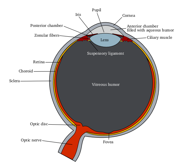
English: Schematic diagram of the human eye
|
|||||
| المصدر |
Own work using: |
|||||
| المؤلف | Delta G | |||||
| إصدارات أخرى |
[] All eye
By languages
For translate
Anterior segment
|


ترخيص
| هذا الملفُّ مُرخَّص بموجب رخصة المشاع الإبداعي نسبة المُصنَّف إِلى مُؤَلِّفه - المشاركة بالمثل 3.0 العامة Subject to disclaimers. | ||
| ||
| تمت إضافة علامة الترخيص لهذا الملف كجزء من رخصة جنو للوثائق الحرة تحديث الترخيص. |

|
يسمح نسخ وتوزيع و/أو تعديل هذه الوثيقة تحت شروط رخصة جنو للوثائق الحرة، الإصدار 1.2 أو أي إصدار لاحق تنشره مؤسسة البرمجيات الحرة؛ دون أقسام ثابتة ودون نصوص أغلفة أمامية ودون نصوص أغلفة خلفية. نسخة من الرخصة تم تضمينها في القسم المسمى GNU Free Documentation License. Subject to disclaimers. |
Eye Anatomy
A guide to the many parts of the human eye and how they function.
The ability to see is dependent on the actions of several structures in and around the eyeball. The graphic below lists many of the essential components of the eye's optical system.
When you look at an object, light rays are reflected from the object to the cornea, which is where the miracle begins. The light rays are bent, refracted and focused by the cornea, lens, and vitreous. The lens' job is to make sure the rays come to a sharp focus on the retina. The resulting image on the retina is upside-down. Here at the retina, the light rays are converted to electrical impulses which are then transmitted through the optic nerve, to the brain, where the image is translated and perceived in an upright position!
Think of the eye as a camera. A camera needs a lens and a film to produce an image. In the same way, the eyeball needs a lens (cornea, crystalline lens, vitreous) to refract, or focus the light and a film (retina) on which to focus the rays. If any one or more of these components is not functioning correctly, the result is a poor picture. The retina represents the film in our camera. It captures the image and sends it to the brain to be developed. The macula is the highly sensitive area of the retina. The macula is responsible for our critical focusing vision. It is the part of the retina most used. We use our macula to read or to stare intently at an object.
source: http://www.stlukeseye.com/Anatomy.asp
تاريخ الملف
اضغط على زمن/تاريخ لرؤية الملف كما بدا في هذا الزمن.
| زمن/تاريخ | صورة مصغرة | الأبعاد | مستخدم | تعليق | |
|---|---|---|---|---|---|
| حالي | 12:00، 5 يونيو 2006 | | 600 × 550 (54 كيلوبايت) | Eliashc | optimized using optipng. |
| 05:37، 14 مارس 2005 |  | 600 × 550 (73 كيلوبايت) | Delta G | Schematic diagram of the human eye(zonule fibers -> zonular fibers(more common)) | |
| 04:58، 14 مارس 2005 |  | 600 × 550 (72 كيلوبايت) | Delta G | Schematic diagram of the human eye(smaller, vitreous fluid -> vitreous humor) | |
| 04:36، 14 مارس 2005 |  | 866 × 793 (115 كيلوبايت) | Delta G | Schematic diagram of the human eye |
استخدام الملف
الصفحتان التاليتان تستخدمان هذا الملف:
الاستخدام العالمي للملف
الويكيات الأخرى التالية تستخدم هذا الملف:
- الاستخدام في en.wikipedia.org
- الاستخدام في fa.wikipedia.org