
ملف:Spindle centriole - embryonic brain mouse - TEM.jpg

الملف الأصلي (1٬283 × 1٬600 بكسل حجم الملف: 901 كيلوبايت، نوع MIME: image/jpeg)
| هذا ملف من ويكيميديا كومنز. معلومات من صفحة وصفه مبينة في الأسفل. كومنز مستودع ملفات ميديا ذو رخصة حرة. |
ملخص
| الوصف |
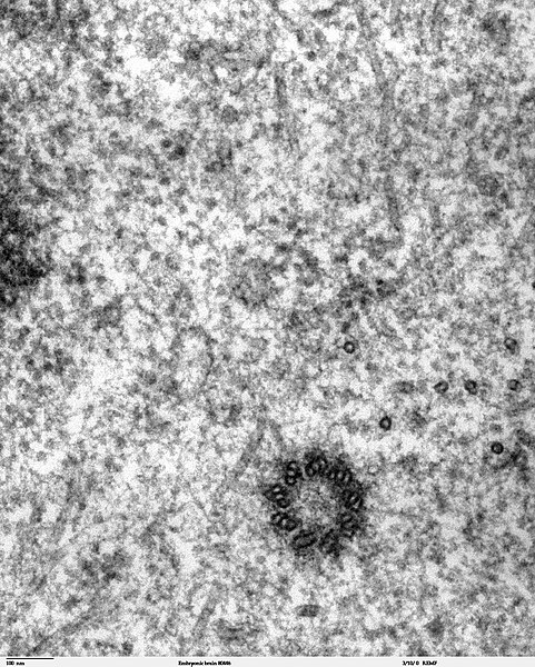
Transmission electron microscope image of a thin section cut through the developing brain tissue (telencephalic hemisphere) of an 11.5 day mouse embryo. This high magnification image of "Embryonic brain 80445" show a spindle centriole and some spindle microtubules visible in the cytoplasm of a mitotic cell at the luminal surface of the telencephalon. JEOL 100CX TEM References: Marin-Padilla, M. (1985) "Early Vascularization of the Embryonic Cerebral Cortex: Golgi and Electron Microscope Studies", J. Comparative Neurology, 241:237-249 Marin-Padilla, M. and M. Amievo (1989) "Early Neurogenesis of the Mouse Olfactory Nerve: Golgi and Electron Microscope Studies", J. Comparative Neurology, 288:339-352 |
| المصدر | |
| المؤلف | Louisa Howard, Miguel Marin-Padilla |
| الترخيص (إعادة استخدام هذا الملف) |
PD |
ترخيص
| وضع -Louisa Howard, Miguel Marin-Padilla-، وهو المؤلف، هذا العمل في النِّطاق العامِّ. يسري ذلك في كل أرجاء العالم. في بعض البلدان، قد يكون هذا التَّرخيص غيرَ مُمكنٍ قانونيَّاً، في هذه الحالة: يمنح Louisa Howard, Miguel Marin-Padilla الجميع حق استخدام هذا العمل لأي غرض دون أي شرط ما لم يفرض القانون شروطًا إضافية.
|
تاريخ الملف
اضغط على زمن/تاريخ لرؤية الملف كما بدا في هذا الزمن.
| زمن/تاريخ | صورة مصغرة | الأبعاد | مستخدم | تعليق | |
|---|---|---|---|---|---|
| حالي | 22:02، 2 نوفمبر 2006 | | 1٬283 × 1٬600 (901 كيلوبايت) | Patho | {{Information |Description=Transmission electron microscope image of a thin section cut through the developing brain tissue (telencephalic hemisphere) of an 11.5 day mouse embryo. This high magnification image of "Embryonic brain 80445" show a spindle cen |
استخدام الملف
الصفحة التالية تستخدم هذا الملف:
الاستخدام العالمي للملف
الويكيات الأخرى التالية تستخدم هذا الملف:
- الاستخدام في bg.wikipedia.org
- الاستخدام في bs.wikipedia.org
- الاستخدام في ca.wikipedia.org
- الاستخدام في cs.wikipedia.org
- الاستخدام في da.wikipedia.org
- الاستخدام في de.wikibooks.org
- الاستخدام في en.wikipedia.org
- الاستخدام في en.wikibooks.org
- الاستخدام في es.wikipedia.org
- الاستخدام في eu.wikipedia.org
- الاستخدام في fr.wikipedia.org
- الاستخدام في gl.wikipedia.org
- الاستخدام في gv.wikipedia.org
- الاستخدام في hu.wikipedia.org
- الاستخدام في kk.wikipedia.org
- الاستخدام في nl.wikipedia.org
- الاستخدام في nl.wikibooks.org
- الاستخدام في pt.wikipedia.org
- الاستخدام في sv.wikipedia.org
- الاستخدام في th.wikipedia.org
- الاستخدام في tr.wikipedia.org